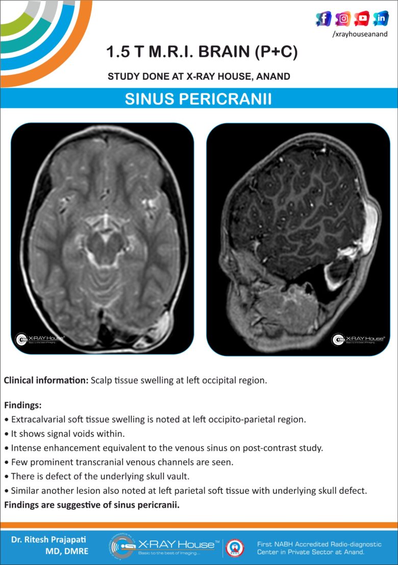
SINUS PERICRANII
Clinical information: Scalp tissue swelling at left occipital region.
Findings:
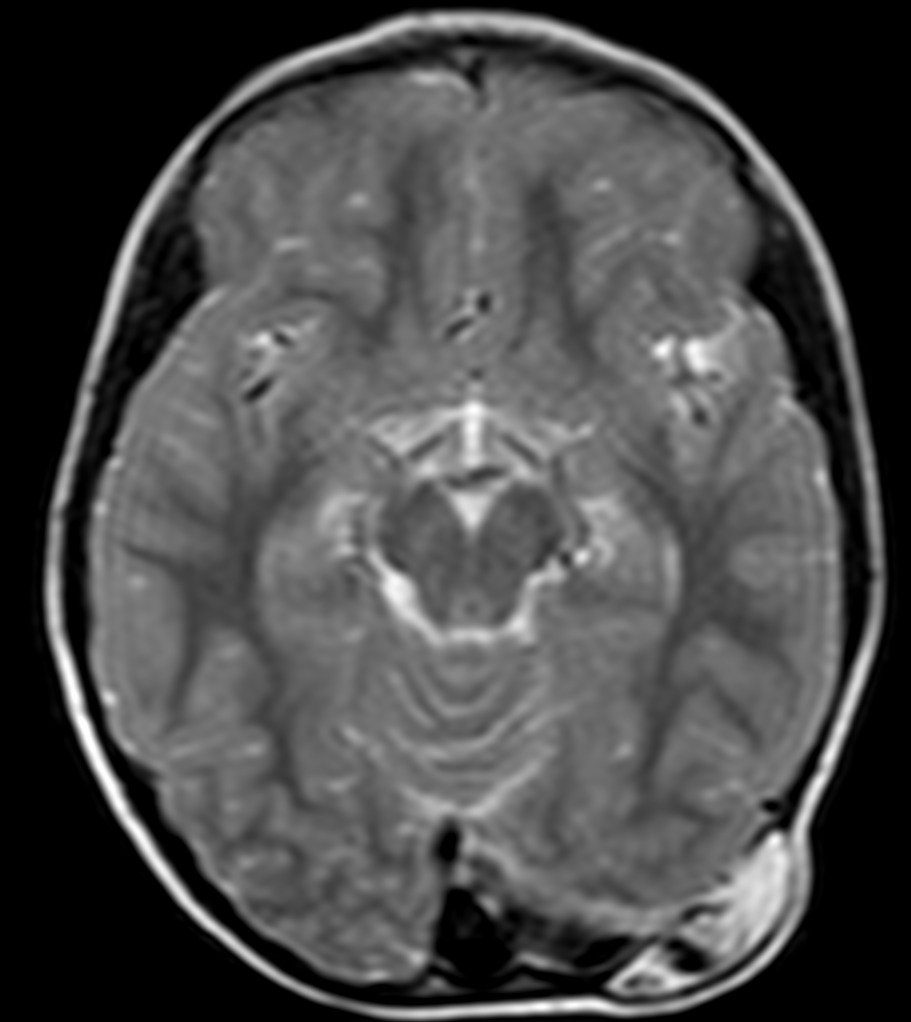
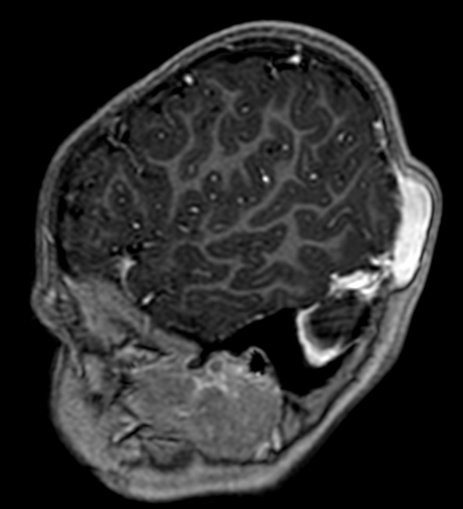
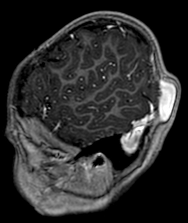
Extracalvarial soft tissue swelling is noted at left occipito-parietal region.
It shows signal voids within.
Intense enhancement equivalent to the venous sinus on post-contrast study.
Few prominent transcranial venous channels are seen.
There is defect of the underlying skull vault.
Similar another lesion also noted at left parietal soft tissue with underlying skull defect.
Findings are suggestive of sinus pericranii.
Discussion:
Sinus pericranii (SP) is a rare disorder characterized by a congenital (or occasionally, acquired) epicranial venous malformation of the scalp.
Sinus pericranii typically presents as soft palpable masses along the midline skull, which may fluctuate in size depending on body positioning.
Sinus pericranii is a venous anomaly where communication between the intracranial dural sinuses and dilated epicranial venous structures exists. That venous anomaly is a collection of non-muscular venous blood vessels adhering tightly to the skull’s outer surface and directly communicating with intracranial venous sinuses through diploic veins. The venous collections receive blood from and drain into the intracranial venous sinuses. The varicosities are intimately associated with the periosteum, are distensible, and vary in size when changes in intracranial pressure occur.
MRI imaging are useful in the detection, detailed evaluation as well as helps in abnormal communication between dural sinuses and the cranial vault.