HEREDITARY MULTIPLE EXOSTOSES


Clinical information: C/o swelling of knee joint.
FINDINGS:
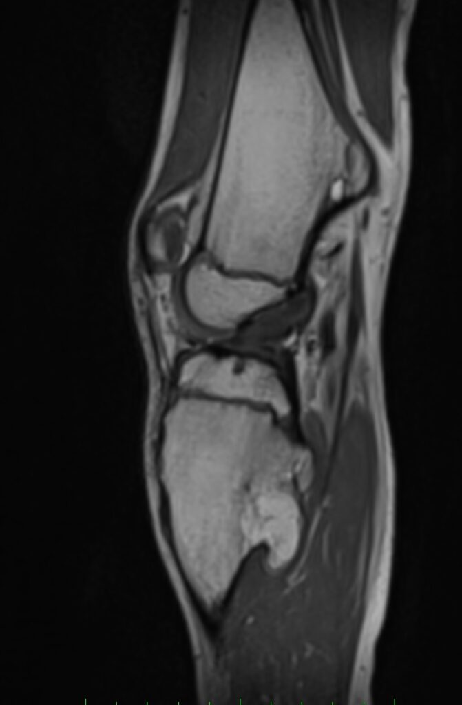
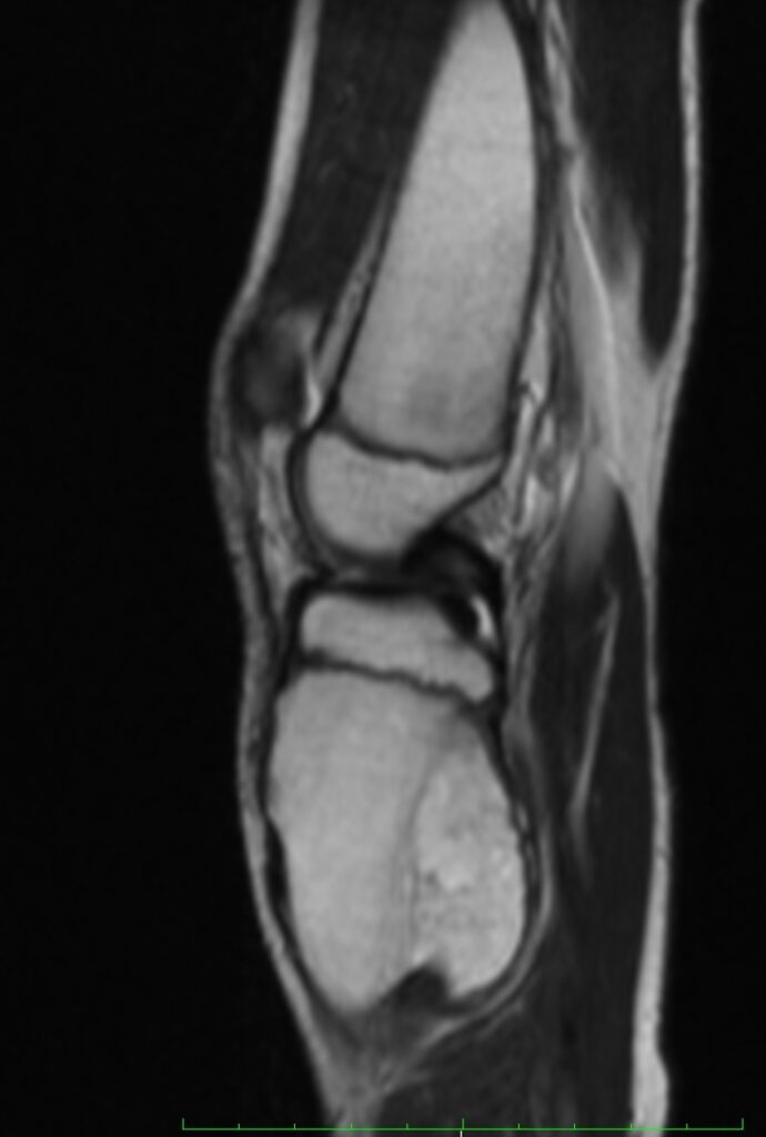
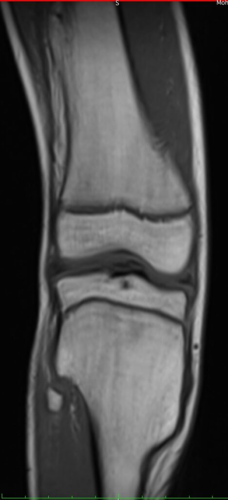
- Bony outgrowths are noted arising from posteromedial aspect of distal shaft of femur. It shows continuity with cortex of femur. No abnormal signal intensity noted within bony outgrowth except few small cystic areas.
- Another similar characteristics bony outgrowths arising from posteromedial aspect of proximal shaft of tibia and anterior aspect of neck of fibula.
- There is expansion of distal meta-diaphysis of femur and proximal meta-diaphysis of tibia.
Above findings are suggestive of hereditary multiple exostosis.
DISCUSSION:
- Hereditary multiple exostoses is a familial disturbance in the growth of cartilaginous bone tissue, most marked at the diaphyso-epiphyseal junction of the long bones.
- It also k/a diaphyseal aclasis, osteochondromatosis.
- It is an autosomal dominant condition, characterized by the development of multiple osteochondromas.
- In rare circumstances, hereditary multiple exostoses occurs together with enchondromatosis in a condition known as ‘metachondromatosis’.
- Most patients are diagnosed by the age of 5 years and virtually all are diagnosed by the age of 12 years. Patients may be asymptomatic with a few small lesions or may be significantly deformed by multiple large osteochondromas.
- This disorder mostly manifest in the tubular bones of the lower extremities. The most common site of involvement seems to be the knees.
- It can also involve any bone in the body except for the calvarium.
- Diagnostic criteria according to the WHO classification of soft tissue and bone tumors (5th edition): Essential: ≥2 radiological osteochondromas at the juxtaepiphyseal region of the long bones and positive family history.
- Complication with this conditions includes – vascular & neural impingement, fracture, deformity, bursitis, ankylosis and rarely malignant transformation.
- Diagnosis is based on the typical radiographic findings of multiple exostoses with associated skeletal deformities.
- Plain radiographs of the affected region remain the mainstay of radiological diagnosis in HME helping to readily identify exostoses and bony deformities.
- However, MRI imaging are useful in the detection, detailed evaluation as well as helps in identifying the complications of HME.
Article Tags:
3.0 T · MRI
Article Categories:
1.5 MRI
Likes:
0 


