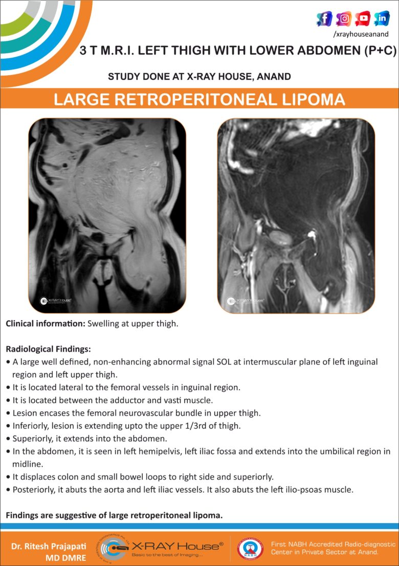
LARGE RETROPERITONEAL LIPOMA
Clinical information: Swelling at upper thigh.
Findings:
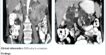
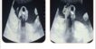
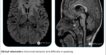
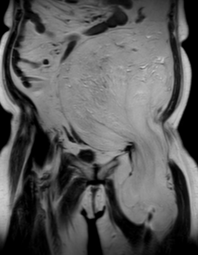
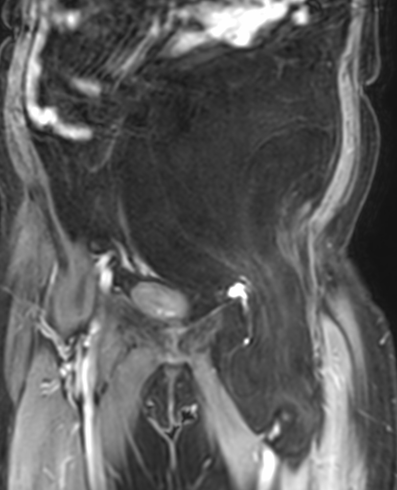
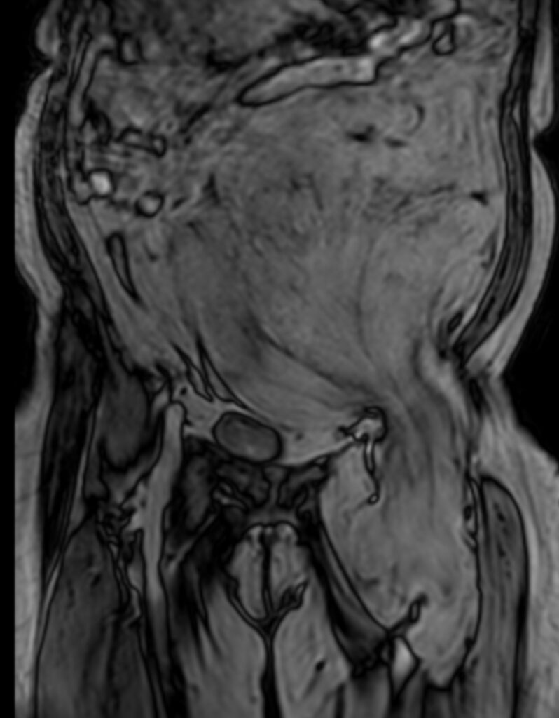
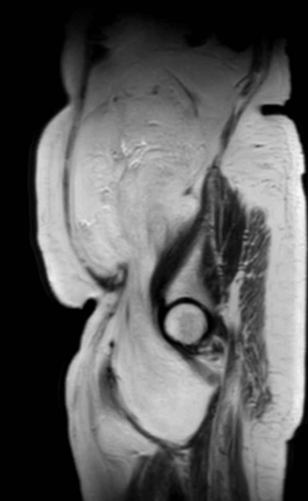
A large well defined, non-enhancing abnormal signal SOL at intermuscular plane of left inguinal region and left upper thigh.
It is located lateral to the femoral vessels in inguinal region.
It is located between the adductor and vasti muscle.
Lesion encases the femoral neurovascular bundle in upper thigh.
Inferiorly, lesion is extending upto the upper 1/3rd of thigh.
Superiorly, it extends into the abdomen.
In the abdomen, it is seen in left hemipelvis, left iliac fossa and extends into the umbilical region in midline.
It displaces colon and small bowel loops to right side and superiorly.
Posteriorly, it abuts the aorta and left iliac vessels. It also abuts the left ilio-psoas muscle.
Findings are suggestive of large retroperitoneal lipoma.
Discussion:
- Retroperitoneal lipoma is benign proliferation and collection of mature fat cells. It is the most frequent soft tissue tumor in adults.
- Lipomas are classified according to the morphologic characteristics into fibrolipoma, conventional lipoma, angiolipoma, myelolipoma, spindle cell lipoma, and myelolipoma.
- They are ordinarily occupying the subdermal tissues of the extremities and trunk.
- Occurrence of lipoma in the retroperitoneal region is an extremely rare finding. In fact, all of the primary retroperitoneal tumors account for only 0.2% of whole body neoplasms.
- Retroperitoneal lipoma may arise from the adipose, connective, muscle, lymphatic or nerve tissues, or it may originate from the mesentery, Gerota’s fascia, or urogenital tract.
- MRI imaging are useful in the detection and detailed evaluation of retroperitoneal lipoma.