SPONTANEOUS SPLENIC RUPTURE


Clinical information: Abdominal pain & vomiting.
FINDINGS:
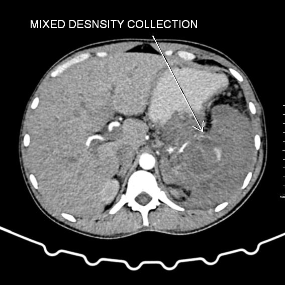
- Spleen is enlarged in size.
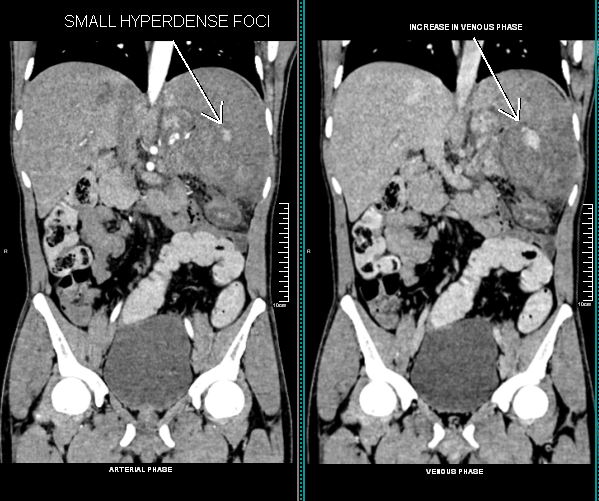
- Mixed density collection in splenic parenchyma extends into the splenic hilum.
- Small hyperdense focus within the collection is seen on the arterial phase, which further increases in size as seen on the delayed scans.
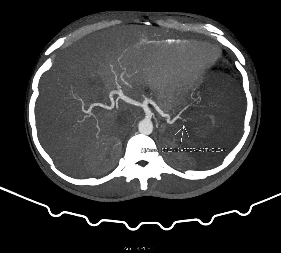
- One of the branches of the splenic artery at the mid-pole is noted adjacent to an active leak.
- Adjacent splenic parenchyma at mid and lower pole region appears hypo-attenuated.
Above findings suggest possibility of spontaneous splenic rupture with active leak (?) from one of the branches of splenic artery.
DISCUSSION:
- Splenic rupture is mainly caused by trauma. But in some rare cases, it can occur without obvious trauma, known as “atraumatic splenic rupture” or “spontaneous spleen rupture”.
- It is a life-threatening diagnostic and therapeutic emergency.
- Non-traumatic ruptures of the spleen can occur in 1 or 2 phases.
- Single stage rupture manifests as an acute pattern rapidly progressing to hemorrhagic shock.
- Two-stage rupture, a subcapsular hematoma is first formed. The symptomatology is sub-acute combining episodes of epigastric or left hypochondrium pain radiating to the left shoulder.
- The spleen is an immunological organ affected by hematological and non-hematological diseases. Spontaneous spleen rupture most likely occurred due to infectious causes, hematological diseases and neoplastic disorders.
- Patients with spontaneous splenic rupture usually present with abdominal pain, signs of hypovolemic shock and tenderness in the left upper quadrant. Nausea and vomiting, syncope and vertigo also reported among clinical symptoms.
- The clinical presentation is not specific and may be confused with other surgical emergencies.
- Ultrasound and CECT – Abdomen scans are used to establish the diagnosis.
- However, CECT scan is the modality of choice for diagnosis and evaluation.
Article Categories:
CT SCAN
Likes:
0 


