RABIES ENCEPHALOMYELITIS


CLINICAL INFORMATION: H/o Dog bite.
FINDINGS:
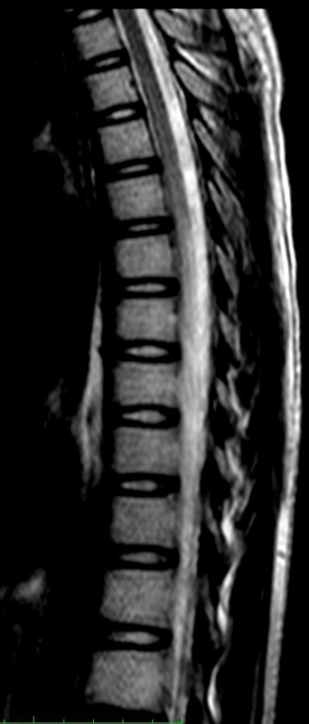
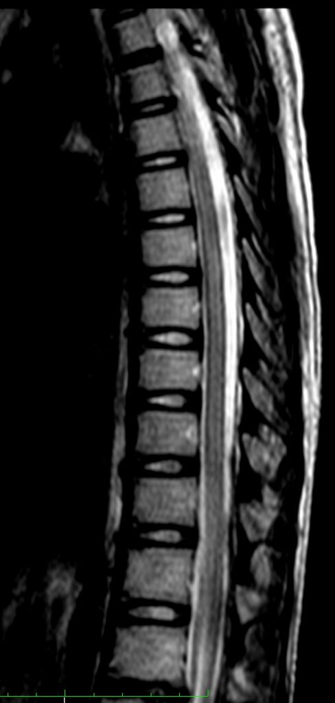
- Diffuse hyperintense signal in the dorsal spinal cord extending from D1 to L1 level.
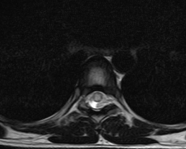
- Hyperintensity involves whole of the cross section of the spinal cord.
- Spinal cord appears slightly bulky.
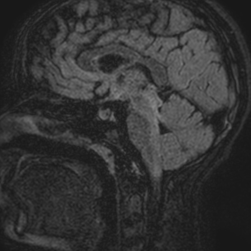
- FLAIR & T2W hyperintensity noted in the dorsal aspect of midbrain, pons and medulla oblongata and cervico-medullary junction.
- No abnormal enhancement noted on post-contrast study.
Findings suggest possibility of Rabies encephalomyelitis.
DISCUSSION:
- Rabies encephalitis is a rapidly progressive CNS infection resulting from infection by a member of an RNA virus of the family Rhabdoviridae, genus Lyssavirus, most commonly transmitted to humans, from infected animal vectors, via a bite.
- It results in rapid neurological deterioration and in almost all instances progresses to death.
- Typically there is an incubation period of 2 weeks to 2 months.
- The main imaging feature is an increase in T2/FLAIR signal in the affected parts of the brain and spinal cord, with a predilection for grey matter structures. As the disease progresses, swelling becomes more marked and petechial hemorrhages occur.
- In classic rabies encephalitis, increased T2 signal has a predilection for the basal ganglia, thalami, hypothalami, brainstem, limbic system, and spinal cord as well as the frontal and parietal lobes. In paralytic rabies, the involvement of the spinal cord and medulla is more pronounced.
MRI Brain and spinal cord is the only modality of any use in the diagnosis of CNS rabies, as CT is usually normal.
Article Tags:
BRAIN · L.S.SPINE · MRI
Article Categories:
1.5 MRI
Likes:
0 


