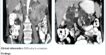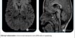
Congenital Diaphragmatic Hernia
OB – 2/3 Trimester Scan Report (23-24 wks)
Findings :
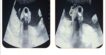
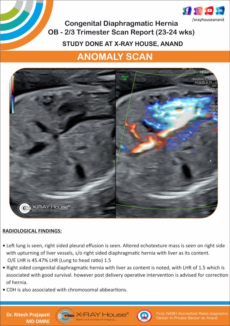
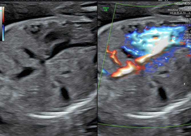
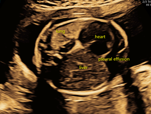
Left lung is seen, right sided pleural effusion is seen. Altered echotexture mass is seen on right side with upturning of liver vessels, s/o right sided diaphragmatic hernia with liver as its content. O/E LHR is 45.47%
LHR (Lung to head ratio) 1.5
Right sided congenital diaphragmatic hernia with liver as content is noted, with LHR of 1.5 which is associated with good survival. however post delivery operative intervention is advised for correction of hernia.
CDH is also associated with chromosomal abbeartions.
DISCUSSION:
Congenital diaphragmatic herniation (CDH) accounts for a small proportion of all diaphragmatic herniae.
Congenital diaphragmatic hernias are seen in 1 of every 2000-4000 live births. 84% are left-sided, 13% are right-sided and 2% bilateral 6.
Most congenital diaphragmatic hernias are detected either soon after birth or on antenatal ultrasound.
Diaphragmatic development is usually complete by ~9th week of gestation. Congenital diaphragmatic hernias result from failure of fusion of one of the pleuroperitoneal canals at about 8 weeks gestation. They may contain the stomach, intestines, liver, or spleen.
Congenital diaphragmatic herniation can be grouped into two basic types on location:
Antenatal ultrasounds (Fetal Sonography) can demonstrate features that suggest congenital diaphragmatic hernias, such as intrathoracic stomach bubble and mediastinal shift.