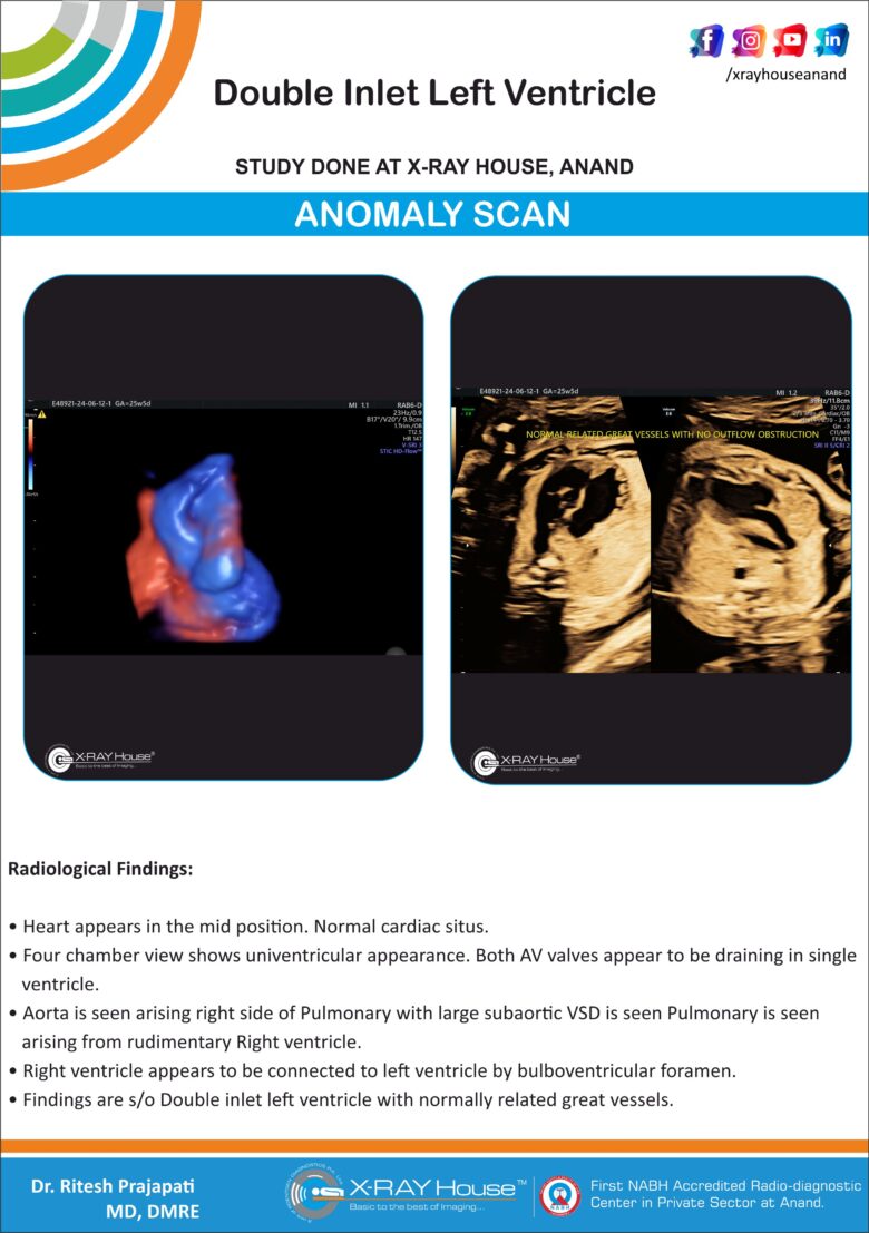
Double Inlet Left Ventricle
Findings :
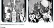
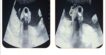
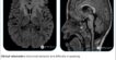
Heart appears in the mid position. Normal cardiac situs.
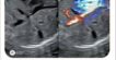
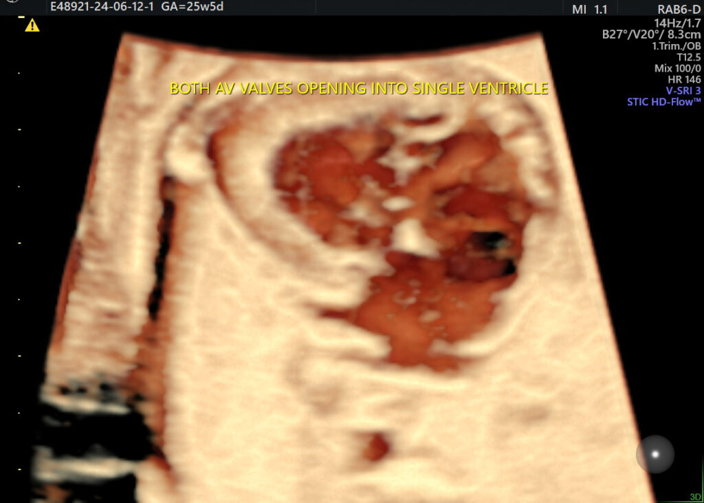
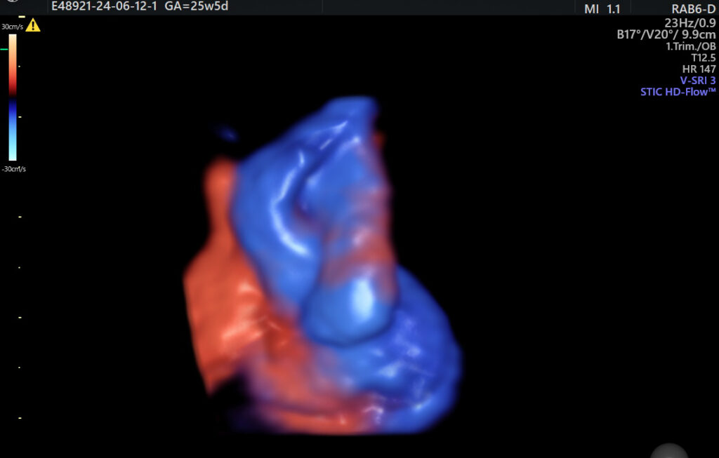
Four chamber view shows univentricular appearance. Both AV valves appear to be draining in single ventricle.
Aorta is seen arising right side of Pulmonary with large subaortic VSD is seen Pulmonary is seen arising from rudimentary Right ventricle.
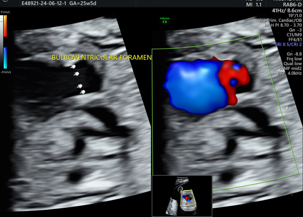
Right ventricle appears to be connected to left ventricle by bulboventricular foramen.
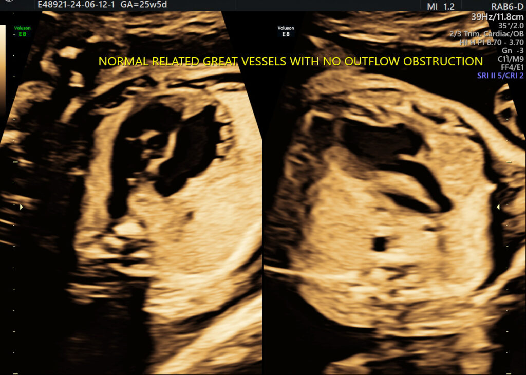
Findings are s/o Double inlet left ventricle with normally related great vessels.
Discussion:
Double Inlet left ventricle (DILV) is a congenital cardiac anomaly with incidence of 0.5 in 1000 live births.
Both the AV valves open into single ventricular chamber most commonly LV. RV is usually small and rudimentary. It receives blood from LV by bubo-ventricular foramen.
Types:
- Normally related vessels with no outflow obstruction.
- Transposed vessels.
- Outlet obstruction.
Antenatal ultrasounds (Fetal Sonography & Fecal echography) can demonstrate features that suggest Double Inlet left ventricle.