CT AORTOGRAM & CORONARY ANGIOGRAM


FINDINGS:
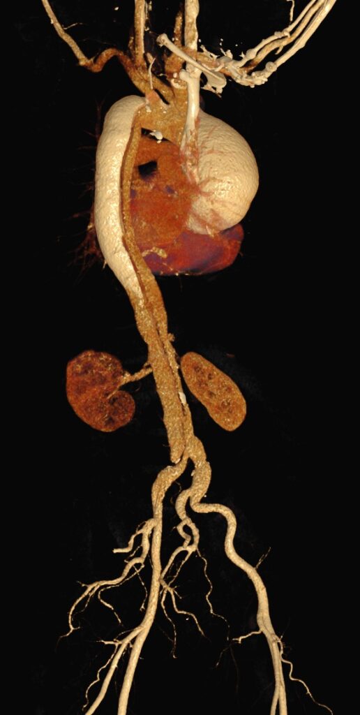
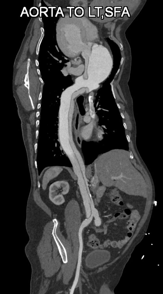
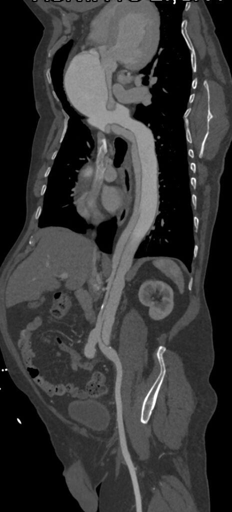
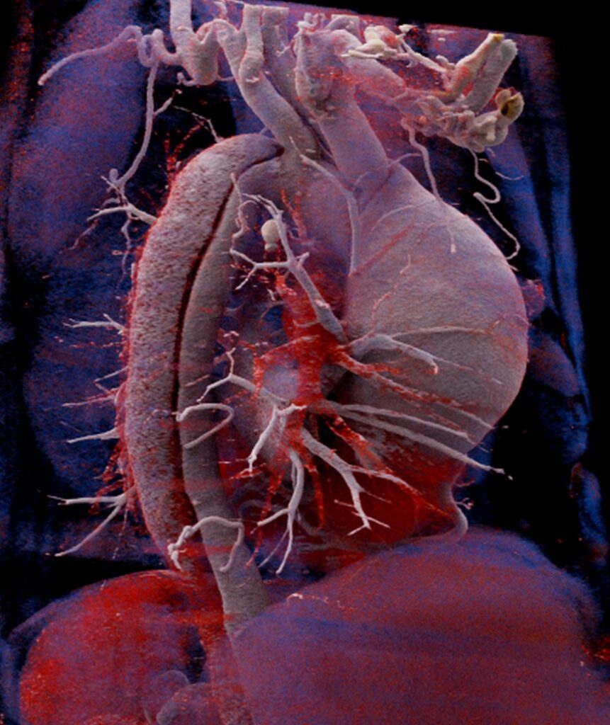
- A dissection flap is seen involving the entire aorta starting from Aortic root (at the level origin of Right Coronary Artery but not involving the origin); involves ascending aorta, arch of aorta, descending thoracic aorta, abdominal aorta, aortic bifurcation and extends till the left common iliac artery; ~ 12-13 mm distal to origin.
- Very small true lumen. Larger false lumen on right antero-lateral aspect of ascending aorta and left postero-lateral aspect of descending aorta without e/o thrombosis in false lumen.
- Aneurysmal dilatation of Ascending aorta and proximal arch of aorta.
- Branches arising from true lumen are: All branches of arch of aorta, celiac artery, SMA and two right sided renal arteries.
- Branches arising from false lumen are IMA and left renal artery.
Above described features are s/o Type ‘A’ Dissection of Aorta associated with Aneurysmal dilatation of Ascending aorta and proximal arch of aorta.
DISCUSSION:
- An aortic dissection is a fatal condition in which a tear occurs in the inner layer of the body’s main artery. Blood rushes through the tear, causing the inner and middle layers of the aorta to split (dissect). If the blood goes through the outside aortic wall, aortic dissection is often deadly.
- Aortic dissections are divided into two groups, depending on which part of the aorta is affected:
- Type A. This more common and dangerous type involves a tear upper aorta (ascending aorta), which may extend into the abdomen.
- Type B. This type involves a tear in the lower aorta only (descending aorta), which may also extend into the abdomen.
- Typical signs and symptoms include sudden severe stomach pain, loss of consciousness, shortness of breath, sudden severe chest pain or upper back pain, leg pain and difficulty walking.
- Aneurysms are focal abnormal dilatation of a blood vessel. They typically occur in arteries & rarely in venous.
- Thoracic aortic aneurysms are relatively uncommon compared to abdominal aortic aneurysms.
- Ascending aortic aneurysms are the most common subtype of thoracic aortic aneurysms.
CT angiography of the aorta is the investigation of choice, as it is used not just for diagnosis and classification but also to rule out any distal complications.
Article Categories:
CT SCAN · Uncategorized
Likes:
0 


