ACRANIA – ANENCEPHALY SEQUENCE

Findings:
Single live intrauterine pregnancy.
Polyhydramnios. (Largest vertical pocket measuring 8.0 cm)
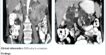
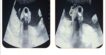
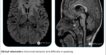
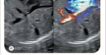
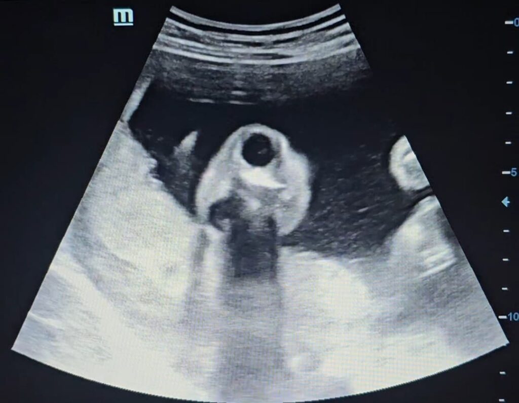
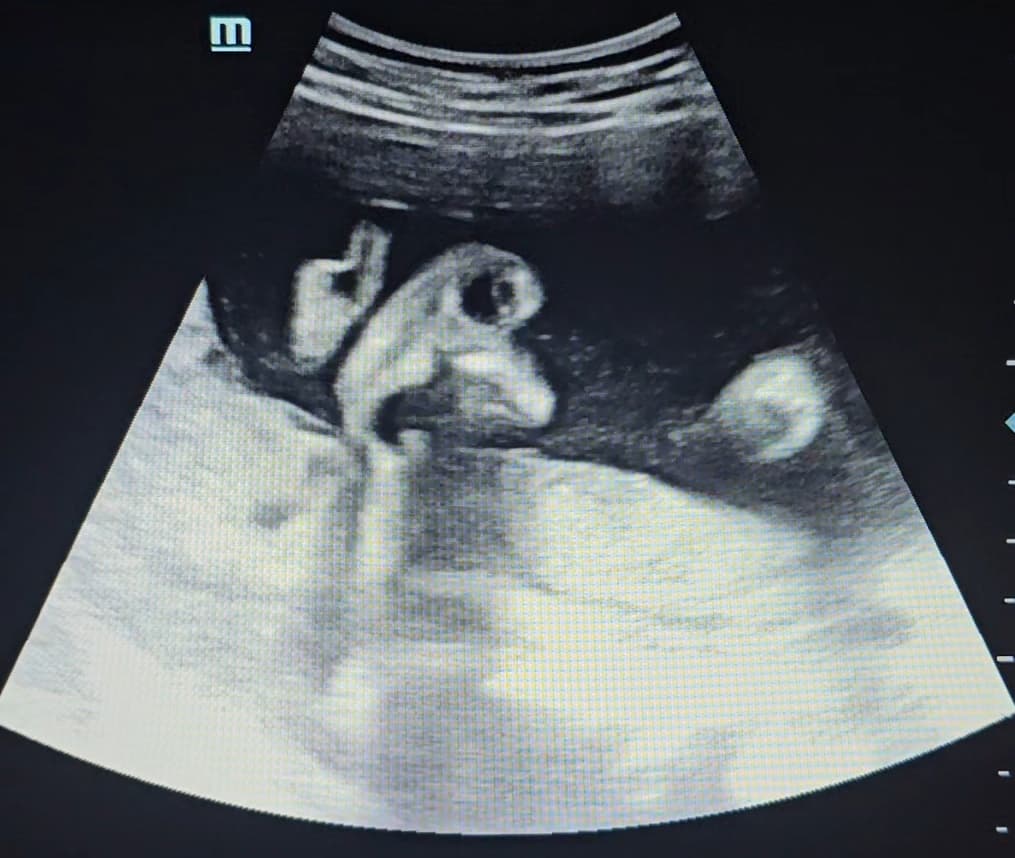
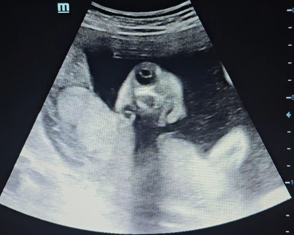
Fetal cranial vault is absent. Bulging of cerebral parenchyma with prominent orbits, — “Frog eye” appearance.
Findings are suggestive of Acrania – anencephaly sequence.
Discussion:
Acrania anencephaly sequence or acrania–exencephaly–anencephaly sequence is the progression from a relatively normal-appearing exposed brain due to an absent cranium (acrania) to an amorphous brain mass (exencephaly) to no recognisable brain tissue (anencephaly).
The acrania anencephaly sequence begins with acrania, which is the most common anomaly affecting the central nervous system with an incidence of ~1:1000 pregnancies.
There are three types of anencephaly and all three are fatal for the fetus:
- Meroanencephaly: The brainstem and midbrain only partially develop. Some skin and skull cover the brain.
- Holoanencephaly: The brain didn’t develop at all. This is the most common type.
- Craniorachischisis: The brain, skull and spine didn’t develop. This is the most severe type.
Signs of anencephaly include:
- High levels of alpha-fetoprotein (a fetal protein) from a blood test or sample of amniotic fluid of the birth mother. This blood test is usually done in the second trimester of pregnancy.
- Too much fluid in the amniotic sac (polyhydramnios) may be seen during a prenatal ultrasound.
- Missing parts of the skull and brain.
- Exposed areas of brain tissue (no skin or skull covering it).
- Smaller head size than expected.